What is corneal topography?
The cornea is the transparent, curved surface at the front of the eye. Corneal topography creates a map of its curvature and shape. The test helps ophthalmologists identify irregularities that may not be obvious during a basic vision check.
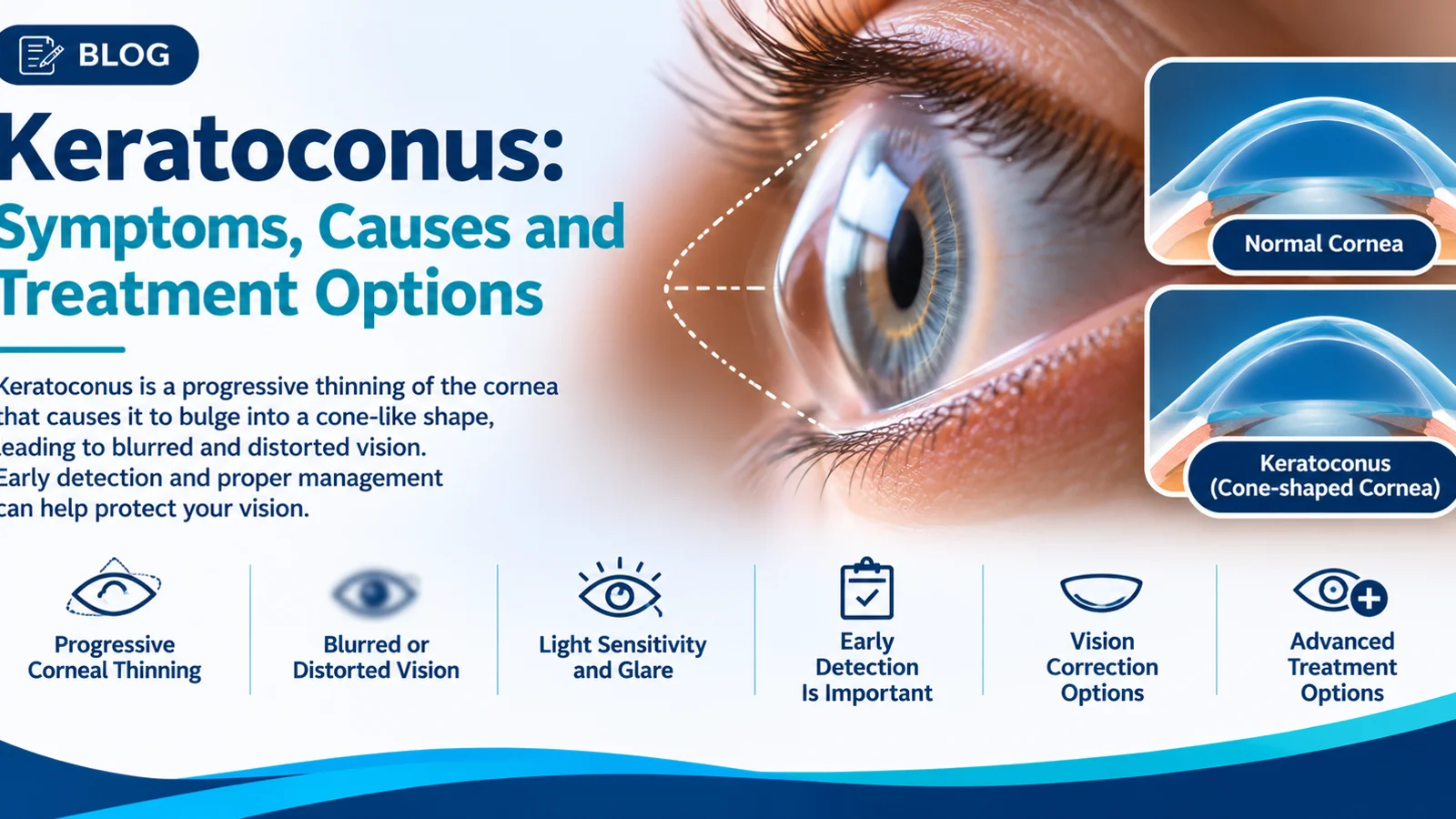
Keratoconus is a condition in which the cornea becomes thinner and develops an irregular, cone-like shape. This can cause blurred or distorted vision, increasing astigmatism and frequent prescription changes.
Why does corneal shape matter?
Ordinary glasses correct many common refractive errors. But when corneal shape becomes irregular, changing the lens power alone may not provide consistently clear vision. A topography map can show an uneven pattern and help the eye specialist decide whether further corneal examination is needed. Other tests may be used to measure thickness and assess the front and back surfaces of the cornea.
What happens during the test?
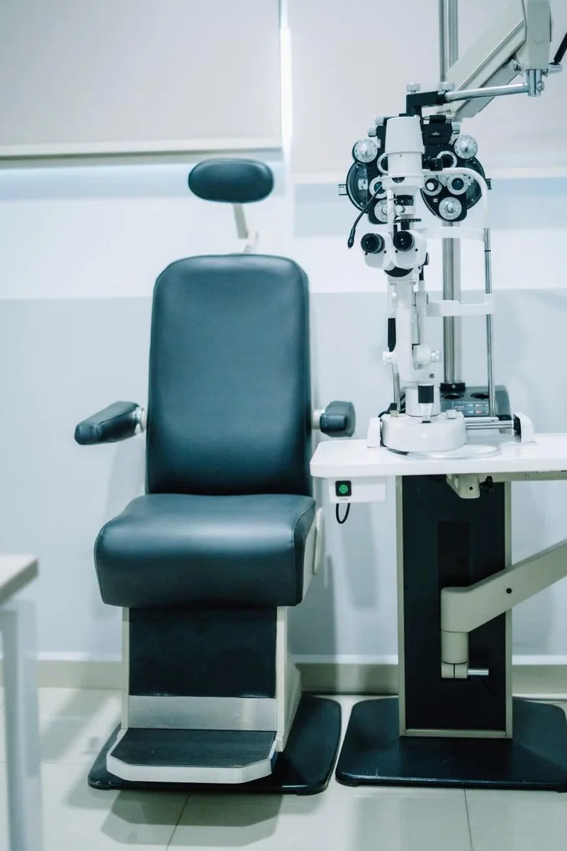
Corneal mapping is generally a quick, non-invasive imaging procedure. You may be asked to look at a target while the device captures images. It is important to follow your clinical team’s instructions, especially if you wear contact lenses, because they can temporarily affect the corneal surface and some measurements.
Can one scan diagnose progression?
A single scan helps establish the cornea’s present shape, but assessment of progression often involves comparing measurements over time along with other clinical information. The doctor may consider age, vision changes, corneal thickness and the reliability of each scan.
How do findings influence care?
For some patients, glasses or specialised contact lenses can improve usable vision. In selected progressive cases, a clinician may discuss corneal cross-linking to try to slow further progression. The appropriate plan depends on the condition and specialist evaluation, and the availability of a particular procedure should be confirmed with the clinic.
Keratoconus evaluation in Indore
Shanti EyeTech describes corneal assessment and imaging as part of its keratoconus care. If your power changes frequently or vision stays distorted despite updated spectacles, arrange an examination rather than assuming another pair of glasses will solve the problem.
Next step
If you have questions about your vision or are considering an eye examination, contact Shanti EyeTech in Indore to discuss an appropriate evaluation. A consultation and examination are needed before any diagnosis or treatment decision.
Medical information is provided for general education and does not replace individual professional advice. Seek urgent ophthalmic care for sudden loss of vision, severe eye pain, or other acute symptoms.
Frequently Asked Questions
Is corneal topography the same as a spectacle power test?
No. Topography maps corneal shape; refraction determines how lenses can improve vision.
Does the scan hurt?
Corneal topography is usually non-contact and not painful.
Can keratoconus be diagnosed from symptoms alone?
No. An eye examination and suitable corneal measurements are needed.
Was this article helpful?
The information is for educational purposes and does not replace professional medical advice, diagnosis or treatment.